Gallery
Figures from phantoms and real scans.
Each figure is made by a script in examples/gallery on a laptop GPU (RTX 4070, 8 GB). The synthetic objects are the procedural phantoms in bench/phantoms, voxelised 1.5 times finer than the reconstruction grid, or STL meshes rendered by gVXR, so the data are never made by the projector that inverts them. Errors are relative L2 against the phantom inside the object.
Ruby sphere in a real laminography scan
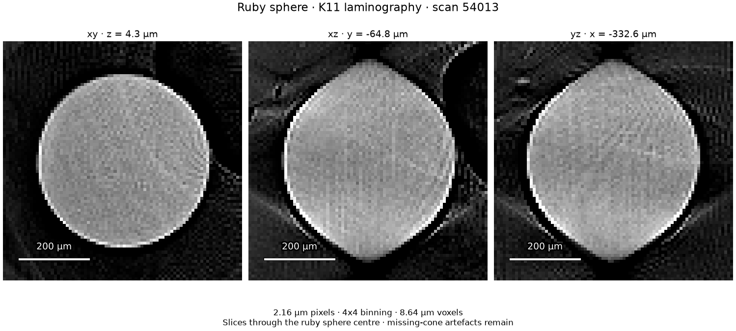
A ruby sphere in Diamond K11 laminography scan 54013. The stored 2.16 µm
pixels are binned 4 × 4 to reconstruct 8.64 µm voxels with the default
tj.align workflow. Slices through the sphere centre share one
grayscale range; scale bars are 200 µm. Missing-cone artefacts remain.
uv run python examples/gallery/k11_ruby_sphere.py k11-54013.nxs k11-ruby-sphere.pngGlass spheres between PEKK plates
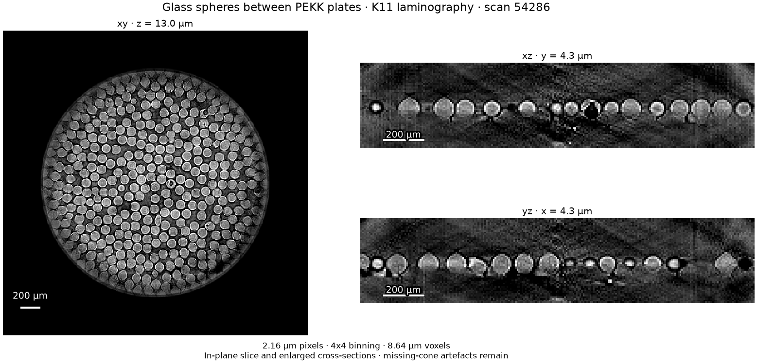
Glass spheres between PEKK plates in Diamond K11 laminography scan 54286.
The stored 2.16 µm pixels are binned 4 × 4 to reconstruct 8.64 µm voxels with
the default tj.align workflow. An in-plane slice through the layer is shown
beside two enlarged cross-sections; scale bars are 200 µm. Missing-cone
artefacts remain.
uv run python examples/gallery/k11_glass_layer.py k11-54286.nxs k11-glass-layer.pngLaminography alignment of a chip package
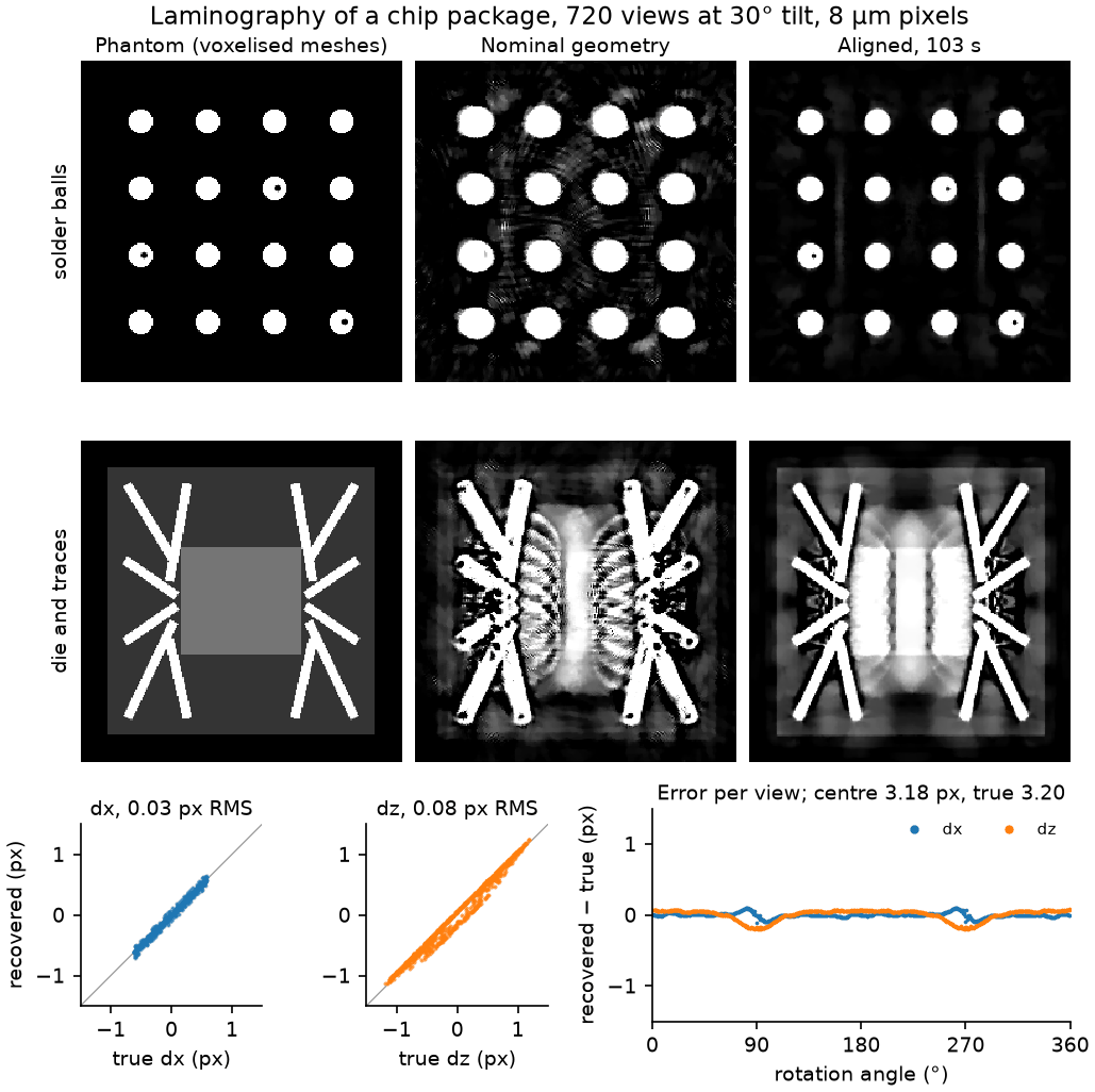
A DIAD-like scan at 25 keV and 30° tilt, rendered by gVXR from the meshes of a
chip package with phase contrast, detector blur, Poisson noise and flat
fields. The detector is 3.2 pixels off the axis and the sample wobbles and
jitters by about two pixels and 0.1° between views. tj.align(scan, mode="cor-then-pose") finds the centre to 0.02 pixels and the per-view shifts
to 0.03 and 0.08 pixels in under two minutes; the voids in the solder balls
reappear. The shifts are compared after removing a rigid motion of the whole
sample, which no reconstruction can detect; the errors that remain are
largest near 90° and 270°. The display is clipped at 1.2 mm⁻¹, so copper and
gold (16 and 85 mm⁻¹) saturate. The die reads about twice its attenuation even
from exact, motionless data: laminography does not measure the slowly varying
part along the sample's normal, so the copper and the layers above and below
bleed into its slice. Slices across the sample plane are left out for the same
reason.
uv run python bench/phantoms/chip_package.py build DATA   # gVXR environment, about 20 minutes
uv run python examples/gallery/chip_laminography.py DATA chip-laminography.png
Ring removal on a real synchrotron scan
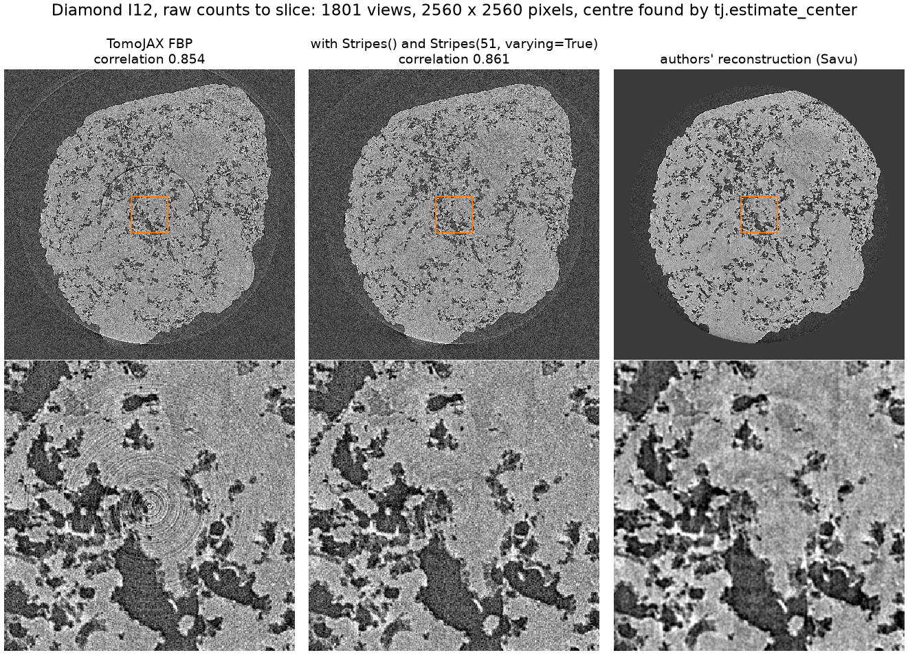
The Diamond I12 scan of Vo et al. (Zenodo 3339629), 1801 views on a
2560-pixel detector, from raw counts. tj.estimate_center places the axis.
Stripes() removes the tight rings round the axis and
Stripes(51, varying=True) the large ring from an intensity-dependent column,
leaving the rock's structure as it was. Correlation with the authors' Savu
slice rises from 0.854 to 0.861.
uv run python examples/gallery/diamond_rings.py full_rows1048-1111.nxs rec_01050.tif diamond-rings.pngFew-view micro-CT of sandstone
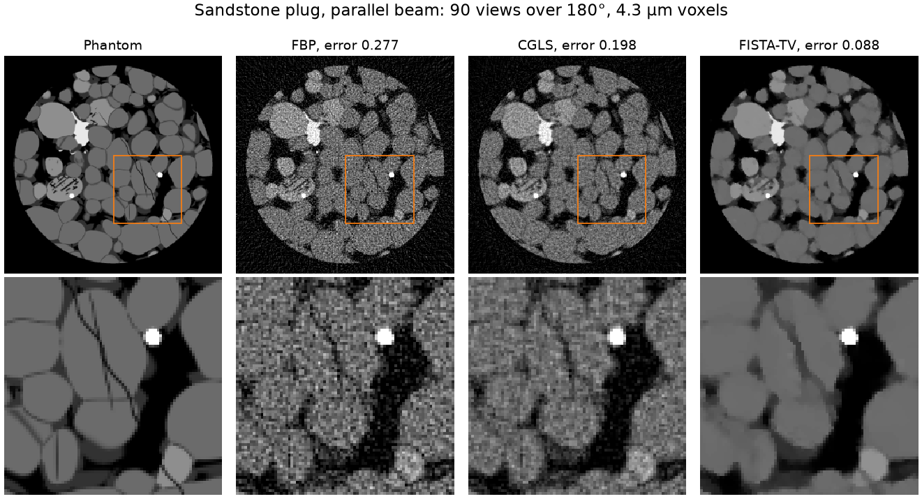
A 1 mm sandstone plug (quartz and feldspar grains, clay seams, pyrite, cracks) in parallel beam with 90 views over 180°. The rock absorbs weakly at 30 keV, so FBP (error 0.277) and 20 CGLS iterations (0.198) are noisy; FISTA-TV with weight 1e-4 (0.088) keeps the grain contacts and cracks.
uv run python examples/gallery/rock_few_views.py rock-few-views.pngA cell wider than the detector
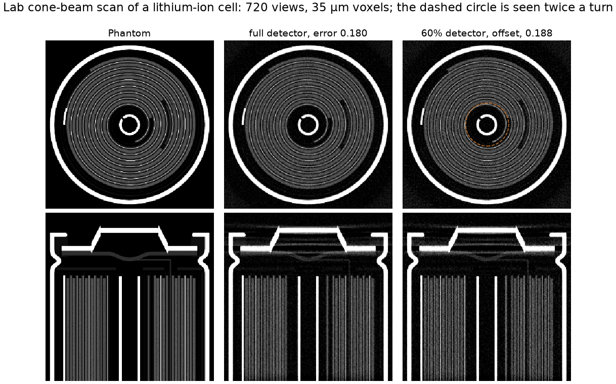
A scaled 18650 lithium-ion cell in lab cone beam at eight times magnification. A detector 60% as wide, offset to see one side of the cell and 1.15 mm past the axis, reconstructs the whole cross-section: FDK weights the rays measured twice a turn (the dashed circle), and the error is 0.188 against 0.180 with a detector covering the cell.
uv run python examples/gallery/battery_offset_detector.py battery-offset-detector.pngWide-cone artefacts on a lattice part
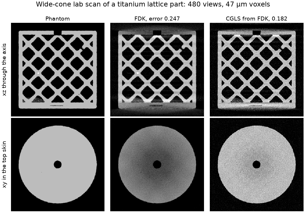
An additively manufactured Ti-6Al-4V octet lattice with pores, a lack-of-fusion flaw and a tungsten inclusion, scanned with a cone of about ten degrees at its top and bottom. FDK's approximation darkens the centre of the top skin and smears the plates (error 0.247); 30 CGLS iterations from FDK fit the cone-beam data and remove it (0.182), at the price of more noise.
uv run python examples/gallery/lattice_cone.py lattice-cone.png